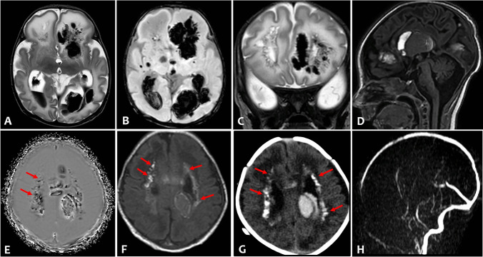
Fig. 1.
Axial T2-weighted (A) and susceptibility weighted imaging (B) of brain MRI showed severe intraventricular hemorrhage and bilateral multiple intraparenchymal hemorrhages, prevailing in the left hemisphere, and enlargement of the posterior horns of the lateral ventricles. Coronal T2-weighted (C) showed multiple cysts in the frontal periventricular white matter. Sagittal T1-weighted (D) revealed also a large intraventricular clot in the III ventricle and hypoplasia of cerebellar vermis. Multiple periventricular calcifications (arrows) were demonstrated on phase axial susceptibility weighted image (E), on T1-weighted (F) image, and on corresponding axial CT (G). There was no evidence of venous thrombosis and vascular malformation on MR venogram (H) and time of flight angiography (not shown)

