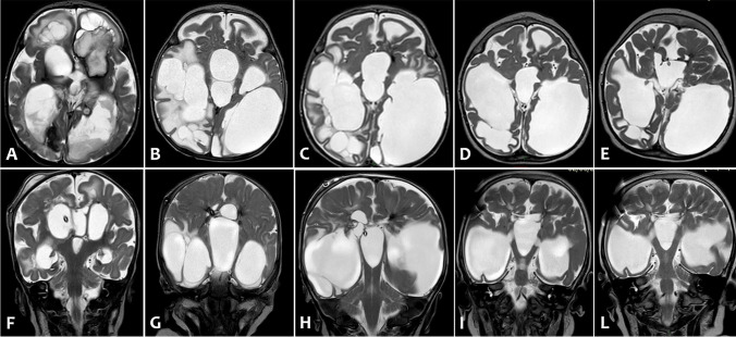
Fig. 4.
Axial (A–E) and coronal (F-L) T2-weighted images of serial brain MRI: (A,F) 1 month of life, (B–G) 3 months of life, (C–H) 4 months of life, (D–I) 15 months of life, (E–L) 2 years of life. Serial MRI scans showed porencephalic cystic evolution and reduced white-matter volume (indicative of destructive changes), and consensual gradual worsening of ventricular dilatation unless a ventriculoperitoneal shunt was implanted at 4 months of life. Last two MRI exams (D-I and E-L) showed a plastic re-organization of porencephalic cysts and lateral ventricles with reduction of signs of cerebrospinal fluid’s decompensation

