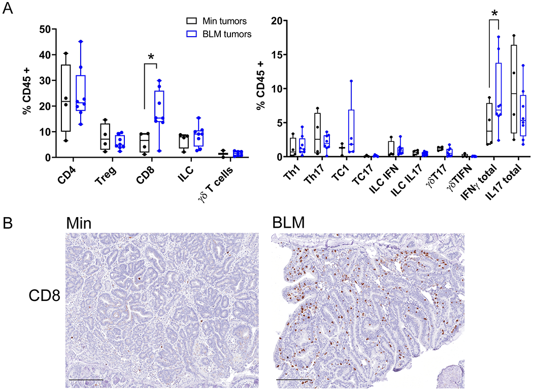
Figure 5. IFN-γ-producing CD8+ cells infiltrate ETBF-induced tumors in BLM mice but not Min mice.

A) Left, Flow cytometry analysis of lamina propria leukocytes isolated from BLM (blue) and Min (black) ETBF-induced colon tumors collected 9-13 weeks post ETBF inoculation. Right, flow cytometry analysis of Intracellular cytokine staining of lamina propria leukocytes isolated from colon tumors in BLM (blue) and Min (black) mice. Each data point represents all tumors for one mouse pooled; Min (N=4) and BLM (N=7) mice from two different experiments. Box limits are set at the third and first quartile range with the central line at the median. *P<0.05 by pair-wise Mann-Whitney U test. B) Representative IHC of CD8+ infiltrating cells in BLM and Min ETBF tumor sections. N= 5-7 mice per group examined and included all tumors along axis of colon. Scale bar, 100μM.
