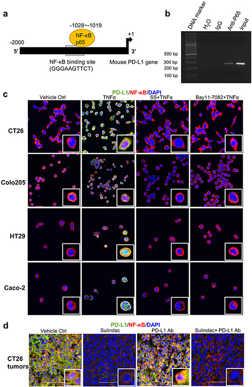
Figure 3: Blockade of NF-κB signaling is involved in sulindac modulation of pMMR/MSS CRC in response to anti-PD-L1 treatment.
(a) Schematic representation of the mouse PD-L1 promoter fragment containing a putative NF-κB p65 binding sequence (GGGAAGTTCT,−1028 to −1019). (b) ChIP assay results showing direct binding of NF- κB p65 to the PD-L1 promoter. CT26 cells were pre-treated with 25 ng/ml TNF-α for 20 min and immunoprecipitated by NF-κB p65 antibody or normal mouse IgG. The isolated DNA fragments were purified from the pull-down complexes and utilized as the templates for PCR amplification. The expected size of the PCR product containing the putative NF-κB p65 binding sequence is 253 bp. Input samples were derived from the starting chromatin that had been used for ChIP. (c) SS attenuates the induction of NF- κB signaling by TNFα. Four CRC cell lines were first treated with 10 μM Bay11-7082 for 2 h and 25 μM SS for 12 h, respectively. DMSO was used as vehicle control. Then, human TNF-α or mouse TNF-α at a concentration of 25 ng/ml was added to these cells and incubated for 20 min. Cells were fixed with 4 % formaldehyde for 10 min and then permeabilized with 1% Triton X-100. After blocking with 1% BSA, the cells were incubated with PD-L1 antibody and NF-κB p65 antibody at 4°C overnight. Then, cells were stained and imaged with confocal microscopy. (d) Sulindac downregulates PD-L1 expression by blocking NF-κB signaling in CT26 tumor issues. The same sample sets for Figure 2 were utilized in this analysis. Red: NF-κB p65; Green: PD- L1; Blue: DAPI. Images were captured by using a Nikon Eclipse Ti2 Laser Confocal Scanning Microscope.

