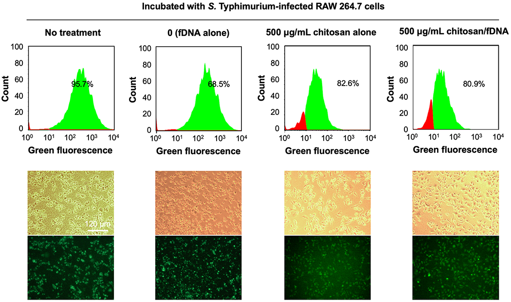
Figure 5.

Intracellular S. Typhimurium infection in RAW 264.7 cells treated with fDNA, chitosan or chitosan/fDNA polyplexes. RAW 264.7 cells were incubated with GFP-expressing S. Typhimurium for 1 h, before overnight treatment. The relative number of RAW 264.7 cells infected with S. Typhimurium was quantified by GFP-expressing cells counted by flow cytometry. Representative bright field and fluorescence micrographs are shown.
