Figure 3.
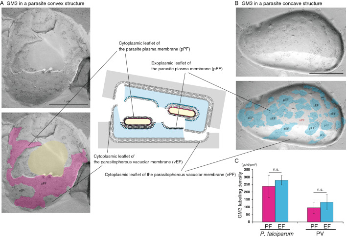
Raft component GM3 is localized in both the EF and the PF of the P. falciparum plasma membrane. In P. falciparum, the labeling of GM3 was detected on both the pPF (A, pink) and pEF (B, blue) leaflets of the plasma membrane. GM3 labeling was also observed on the PV membrane EF (vEF in A) and PV membrane PF (vPF in B). The cytoplasm of P. falciparum is colored with yellow (lower panel in A). Scale bars: 1 μm. (C) Labeling density of GM3 in the P. falciparum membrane and PV membrane in each leaflet. The labeling densities of GM3 on the PF (pink) and the EF (blue) of the P. falciparum plasma membrane and the PV membrane in the erythrocyte are shown. The gold labeling densities on the PF of the parasite plasma membrane and the PV membrane were comparable to those on the EF of each membrane. The mean ± s.e.m. of three independent experiments is shown. The labeling densities of GM3 on the PF were not significantly different (n.s., Student’s t-test) from those on the EF of both the P. falciparum plasma membrane and PV membrane.

