Figure 7.
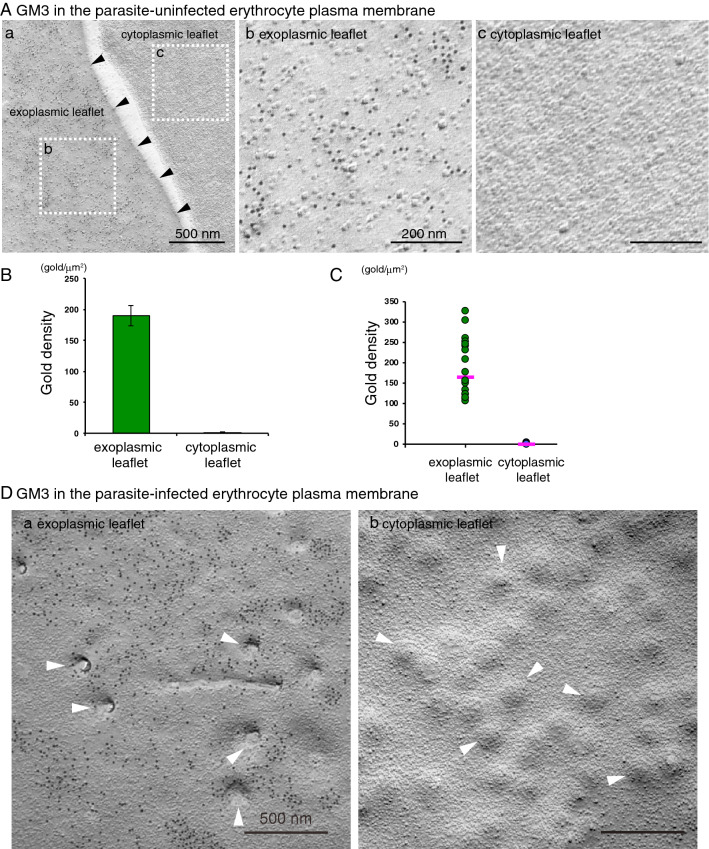
The expression of GM3 in the EF, but not the PF, of the uninfected and infected human erythrocyte plasma membrane. Freeze-fracture replicas of the erythrocyte were labeled by anti-GM3 antibody and colloidal gold (10 nm)-conjugated anti-mouse IgM antibody. (A) In the uninfected erythrocyte, GM3 labeling by 10 nm colloidal gold particles was detected in the EF of the freeze-fractured plasma membrane (a,b). The PF of the plasma membrane was unlabeled (c). The cell boundary is marked by arrowheads (a). The two rectangles in (A) are magnified in (b) (EF) and (c) (PF). The average (B) and scatter diagram (C) of gold labeling density of GM3 on both the PF and the EF of the plasma membrane showed a wide range for each sample on the EF. Pink lines in C indicate the median of each data. (D) In the infected erythrocyte plasma membrane, the knobs were clearly observed as indentations on the EF (arrowheads in a) or protrusions on the PF (arrowheads in b). As in the uninfected erythrocyte plasma membrane, the labeling of GM3 was detected on the EF (a), but not the PF (b) in the infected erythrocyte plasma membrane. Scale bars: 500 nm (Aa,D) and 200 nm (Ab,Ac).

