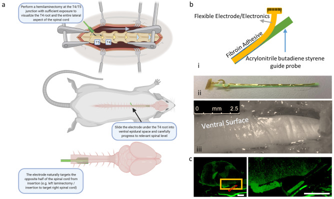
Figure 2.
Surgical approach. (a) We developed a technique to insert highly flexible electronics into the ventral aspect of the cervical rat spinal cord using a dorsal thoracic approach. (b) (i) low bending stiffness biocompatible arrays were fixed to a 3D printed guide probe with biocompatible silk polymer fibroin to (ii) aid in insertion. (iii) Following 2 months of implantation, the array remained positioned at the C5/C6 spinal level lateral to midline. (c) IHC stains for GFAP revealed low bending stiffness arrays do not exhibit enhanced GFAP signal (green) near the spinal surface most proximal to the array, though arrays do appear to become encapsulated in connective tissue over time. Red dashed box—approximate array location, yellow box—location of inset, *all scale bars indicate 500 µm.

