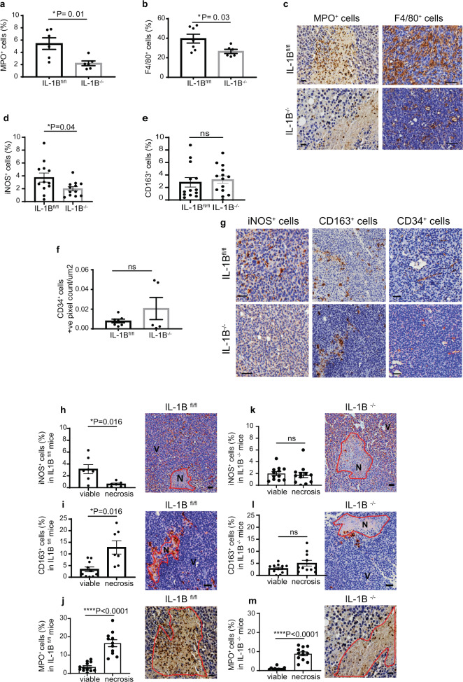
Fig. 2. Microenvironment-derived IL-1B drives the infiltration of innate immune cells with putative anti-tumour function and promotes immune cell subset positioning in primary breast tumours.
a MPO+ neutrophils (*P = 0.01) and b F4/80+ macrophages (*P = 0.03) in the primary tumour of IL-1Bfl/fl and IL-1B−/− mice. Each dot represents a tissue section from a primary tumour isolated from each mouse. c Representative IHC micrographs of MPO and F4/80 staining. d iNOS+ (*P = 0.04), e CD163+ (ns) and f CD34+ (ns) cells in the primary tumour of IL-1Bfl/fl and IL-1B−/− mice. d, e Each dot represents an inner or outer section from a primary tumour obtained from each mouse. f Each dot represents a tissue section from a primary tumour isolated from each mouse. g Representative IHC micrographs of iNOS, CD163 and CD34 staining. Data are mean +/− SEM Two-tailed unpaired t-test with Welch’s correction. Scale bar: 50 µm. h iNOS+ cells in viable and necrotic areas of the primary tumour (tissue section from the tumour core) (P = 0.016) of IL-1Bfl/fl mice determined by IHC. i CD163+ cells in viable and necrotic areas of the primary tumour (P = 0.016) of IL-1Bfl/fl mice determined by IHC. j MPO+ neutrophils in viable and necrotic areas of IL-1Bfl/fl (P < 0.0001). k iNOS+ cells in viable and necrotic regions of primary tumours from IL-1B−/− mice. l CD163+ cells in viable and necrotic areas of primary tumours from IL-1B−/− mice. m MPO+ neutrophils in viable and necrotic areas of IL-1B−/− (P < 0.0001) mice. Data are shown as mean +/− SEM, Two-tailed unpaired t-test with Welch’s correction, ns non-significant. Scale bar: 50 µm in h, i, k, l. Scale bar: 20 µm in j and m.

