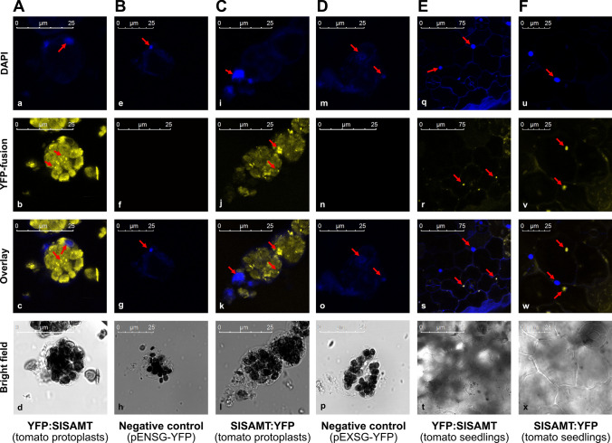
Fig. 4.
Sub-cellular localization of N and C terminal YFP fusions of SlSAMT in tomato protoplasts and seedlings under confocal microscopy. Tomato protoplasts transformed with N terminal (A) and C terminal (C) YFP fusion of SlSAMT showed clear localization in the cytoplasm. Tomato seedlings transformed through the PDS/1000 system with N terminal (E) and C terminal (F) fusion of SlSAMT also showed strong localization in the cytoplasm. DAPI stained cells of each construct which was used as a comparative marker show a clear nuclear signal (a, e, i, m, q,u). Tomato protoplasts transformed with empty plasmid pENSG-YFP used as a negative control for N terminal (B) and pEXSG-YFP as C terminal (D) YFP fusions which did not show any YFP signal (f and n). Arrows highlight the sub-cellular localization of SlSAMT in the cytoplasm while DAPI stained cells in the nucleus. Scale bar 25 μM

