
Figure 4.
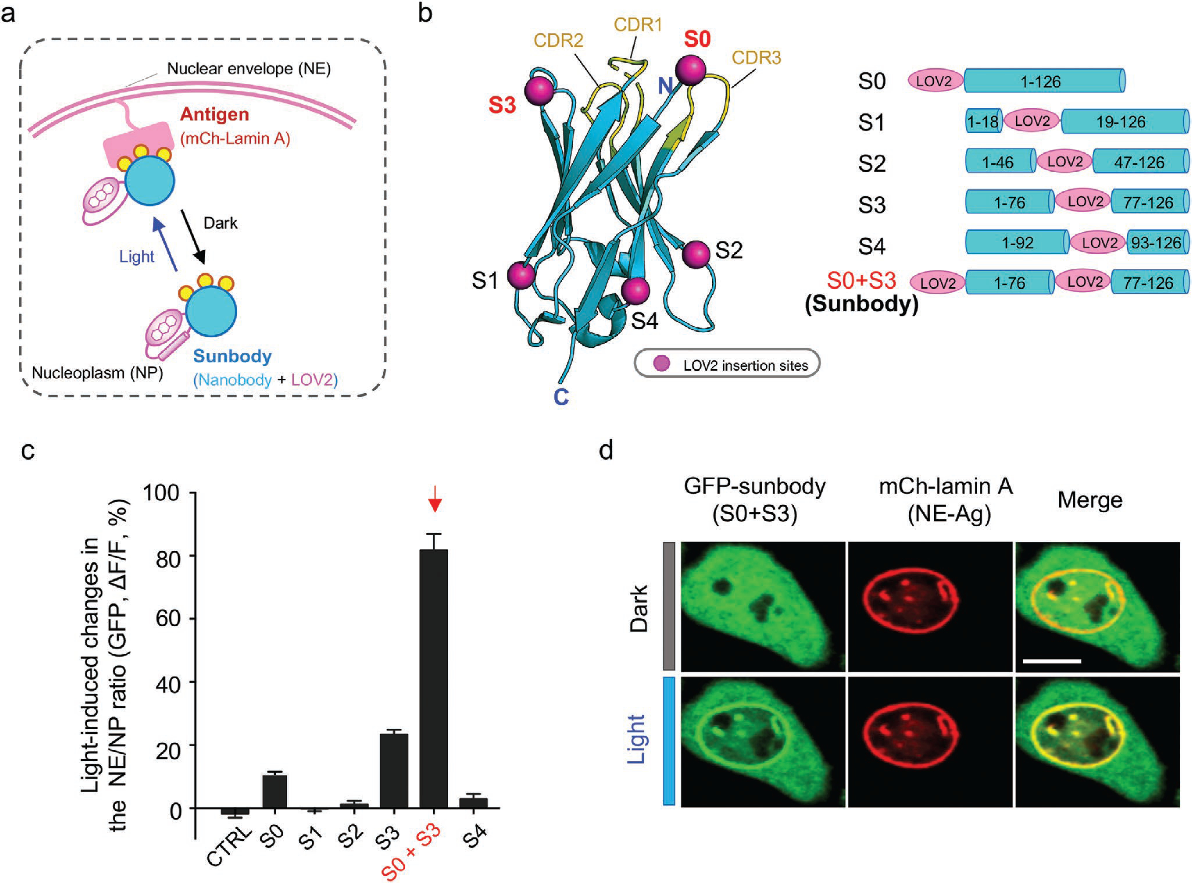
Engineering photoswitchable nanobody (sunbody) to enable light-controllable antigen binding. a) Cartoon depiction of the design and the NP-to-NE translocation assay. Photoswitchable redistribution of an engineered anti-mCherry (mCh) nanobody (designated “sunbody”) is used as the readout. Sunbody is expected to shuttle between NE and NP in a light-dependent manner. Yellow circles represent three CDRs involved in antigen binding. b) Insertion sites for LOV2 mapped to the modeled 3D structure of an anti-mCh nanobody (LaM8). S1, S2, and S4 are located at the opposite side of CDR loops. Both the N-terminus (S0) and S3 are in close proximity to CDRs. See Figure S3, Supporting Information, for detailed sequence information. Data are shown as mean ± SEM. c) Quantification of light-induced changes in the NE/NP ratio for an anti-mCh GFP-tagged sunbody. The combination of LOV2 fusion to the N-terminus (S0) and its additional insertion at S3 led to the strongest light-inducible changes (S0 + S3). See Figure S3, Supporting Information, for light-induced changes of each construct. n = 15–66 cells from three independent assays. Data are shown as mean ± SEM. d) Representative confocal images of a HeLa cell coexpressing sunbody (GFP-tagged LaM8-S3; green) and NE-tethered mCh-lamin A (red) before and after light illumination for 10 s. Scale bar = 10 μm.