
Figure 5.
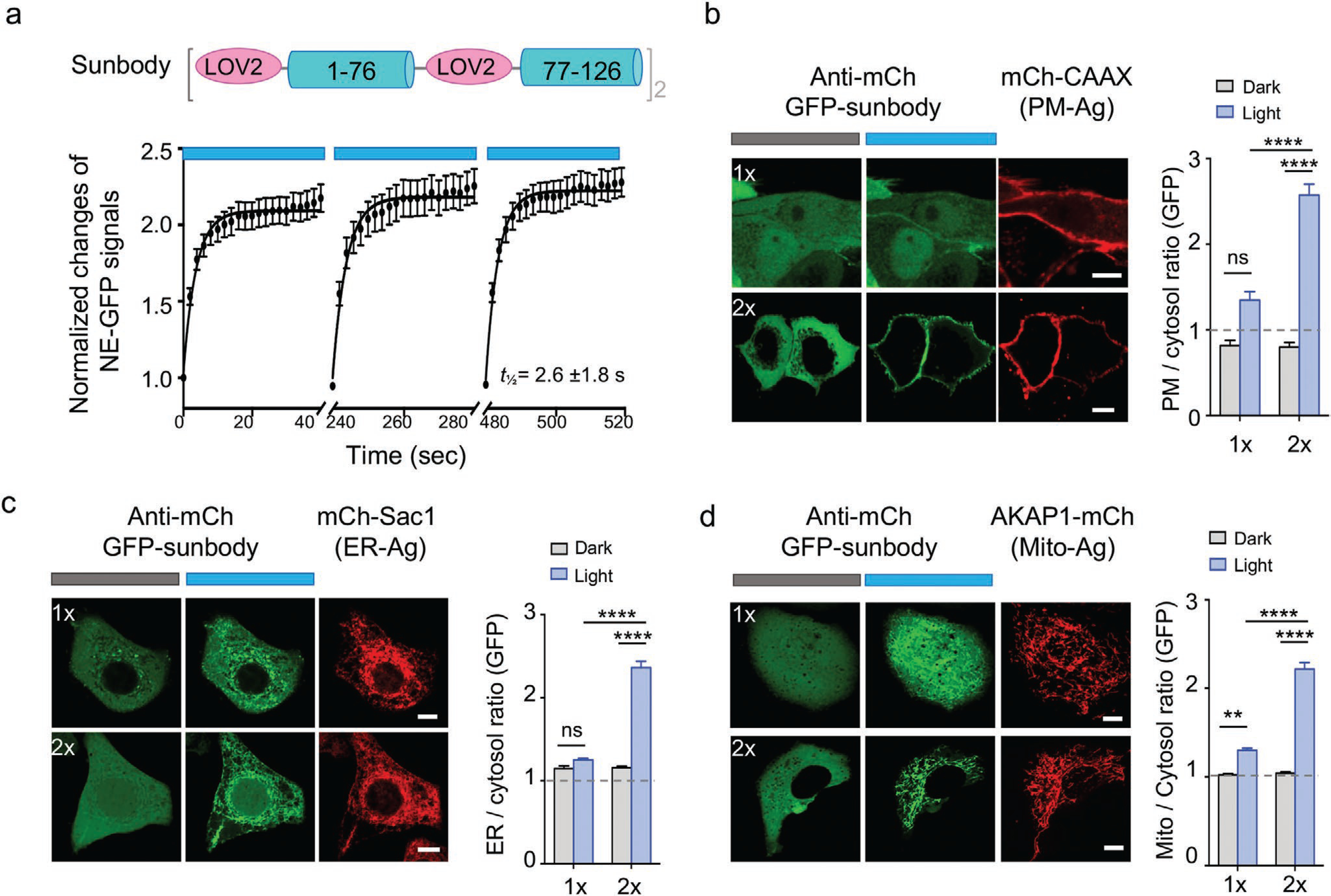
Improved sunbody shows high sensitivity for light-dependent subcellular targeting. Data are shown as mean ± SEM. a) Quantification of the sunbody–antigen interaction in response to three repeated dark–light cycles. The changes in the nucleoplasmic GFP signals were used as the readout. n = 23 cells. b–d) Sunbody used for light-dependent subcellular targeting of its binding partner. HeLa cells were transfected with an anti-mCh GFP-tagged sunbody (1×; green; top panels), or its concatemeric form (2×; green; bottom panels), along with the mCh as antigen (red) tethered to PM (b), ER (c), or outer mitochondrial membrane (d). The quantification of relative GFP signals at the corresponding subcellular organelles before and after light illumination were shown next to the images (n = 15–75 cells). The use of 2xsunbody in a single construct substantially enhanced the signal-to-noise ratio. Scale bar = 10 μm. Also see Movies S3 and S4, Supporting Information. **** p < 0.0001, **p < 0.01 (Two-way ANOVA Tukey’s multiple comparison test).