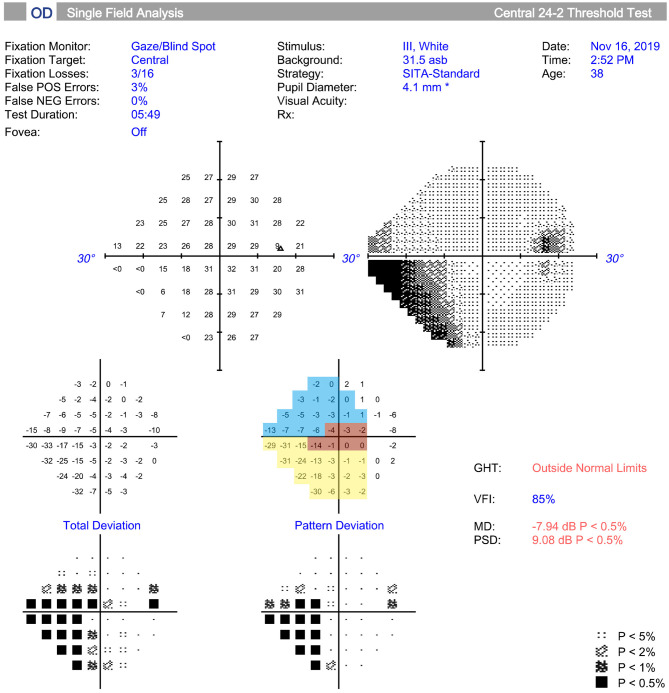
Figure 1.
Measurement and regionalized of the visual field (VF) were demonstrated. The VF was tested (Humphrey automated perimetry SITA 24-2), and regionalized into the temporal (T) (315°-45°, red area), temporal superior (TS) (45°-90°, yellow area), and temporal inferior (TI) (270°-315°, blue area) sectors according to previous study. Regional visual field sensitivity (VFS) was calculated as the sum of the pattern deviations in a specific sector.

