
FIGURE 2.
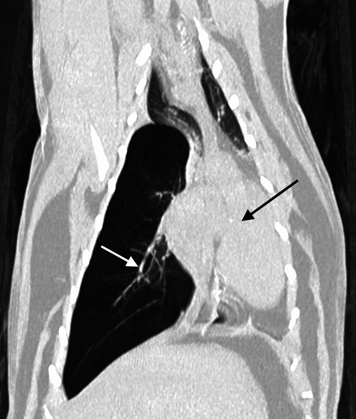
Dorsal computed tomographic image in lung window (WL −500, WW 1400) of the thorax of a cat with lobar emphysema of the right caudal lung lobe. Note the mediastinal shift towards the left (black arrow shows the displaced mediastinal structures). The right caudal pulmonary vessels are thin (white arrow)