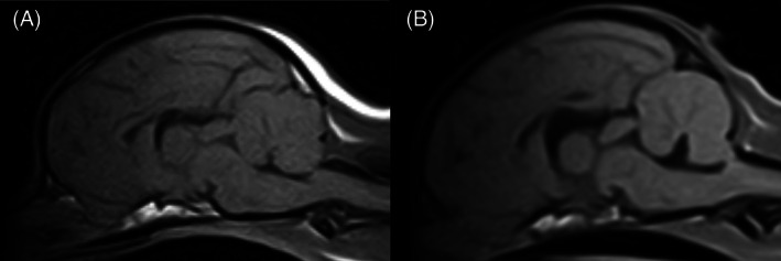
FIGURE 2.

Sagittal T1‐weighted, magnetic resonance image of a Chihuahua with (B) and without (A) fourth ventricle dilatation. A, Chihuahua with a slit‐like fourth ventricle grouped as having no fourth ventricle dilatation. B, Chihuahua with a triangular shaped fourth ventricle and mild deviation of the rostroventral cerebellar tissue grouped as having fourth ventricle dilatation
