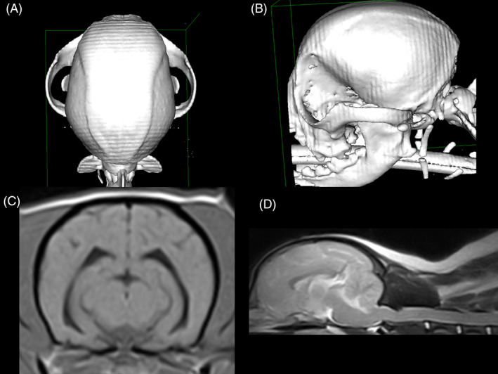
FIGURE 4.

A Chihuahua (weight 4.3 kg) without persistent fontanelles. A,B, Volume‐rendering‐technique computed tomography image of a Chihuahua skull in dorsal (A) and left lateral views (B), showing no persistent fontanelles. C, T1‐weighted transverse brain magnetic resonance image of the same dog from (A, B), lacking ventriculomegaly (ventricular volume 0.57 mm3). D, T2‐weighted sagittal brain and spinal cord (C1‐C5) magnetic resonance image of the same dog from (A, B), showing a very mild medullary elevation, moderate dorsal spinal cord compression at C1‐C2 (sum index 0.43), and lacking syringomyelia
