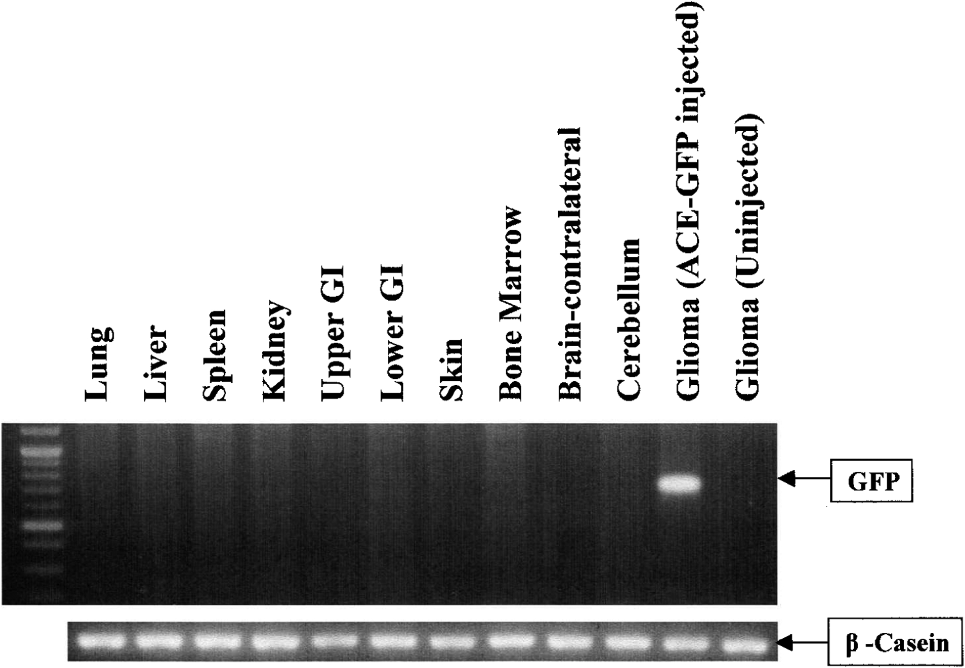
FIG. 4.

RCR biodistribution after intratumoral injection, as determined by genomic PCR analysis. Genomic DNA (0.5 μg) from athymic nu/nu mice with intracranial gliomas injected with ACE-GFP was isolated from U-87 glioma, contralateral supra-tentorial hemisphere (normal brain), bone marrow, skin, lung, liver, spleen, kidney, upper GI tract (esophagus and stomach), and lower GI tract (colon and small intestine) and was analyzed by PCR. The expected size of the full-length GFP product is 700 bp. Only the injected transduced tumor shows a detectable GFP signal. β-Casein gene, a 525-bp fragment, was also amplified as an internal control. Uninjected glioma was used as a negative control.
