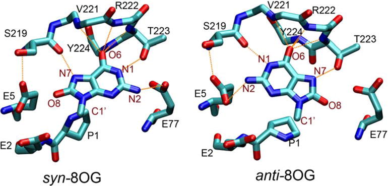
Figure 5.

8OG and surrounding residues in the Fpg-DNA complex with (a, left) syn 8OG as observed in the crystal structure and (b, right) anti 8OG built by rotation around the glycosidic bond of 8OG. Protein residues are labeled in black, and atoms of 8OG are labeled in maroon. Hydrogen bonds are indicated by orange dashed lines. Only the base group of 8OG is shown (the remaining atoms linked to C1′ are not shown).
