Fig. 1.
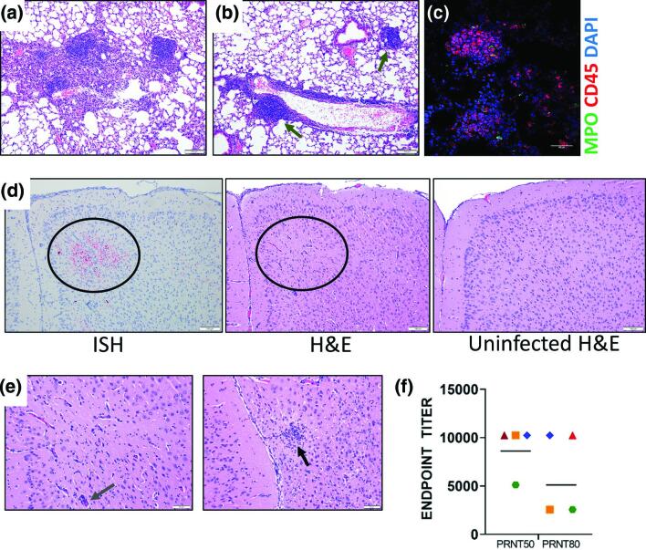
Characterization of K18-hACE2 mouse survivors. (a) H and E staining of mouse lungs showing a focal area of histiocyte infiltration within alveoli and foci of hyperplastic lymphoid tissue from animals that survived SARS-CoV-2 challenge (day 28) (blue arrow). (b) Lung from a surviving mouse showing infiltration of lymphocytes surrounding an intermediate sized vessel, and foci of hyperplastic lymphoid tissue (green arrows). (c) IFA of lung with hyperplastic lymphoid tissue were stained with CD45 and MPO. Cells were counterstained with DAPI to visualized nuclei. (d) Representative ISH staining of infected K18-hACE2 (day 28) mouse demonstrating presence of viral RNA in the cerebral cortex. H and E staining of this same area (circle) shows minimal gliosis and perivascular infiltration of mononuclear leukocytes compared to an uninfected mouse brain in the same region. (e) H and E staining from the same mouse in D showing a focal glial nodule (black arrow) in the rostral cerebral cortex in the image on the right, and a higher magnification of the centre image in panel D showing minimal gliosis and perivascular infiltrates (slate arrow). (f) Mouse serum (n=4 survivors) was evaluated for its ability to neutralize SARS-CoV-2 by plaque reduction assay. Endpoint PRNT50 and PRNT80 titres were plotted from eleven different replicates. The geometric mean titre are plotted, each shape/colour represents an individual mouse (n=4). The limit of quantitation was 40.

