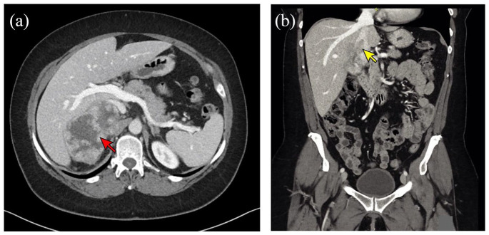
Figure 1.
CT imaging in a patient with advanced ACC. (a) Axial CT image showing a heterogeneously enhancing right adrenal mass (red arrow) measuring 9.9 × 6.0 × 8.0 cm with possible invasion into the liver. (b) Coronal CT image from the same patient showing tumor thrombus within the inferior vena cava (yellow arrow).
ACC, Adrenocortical carcinoma; CT, computed tomography.

