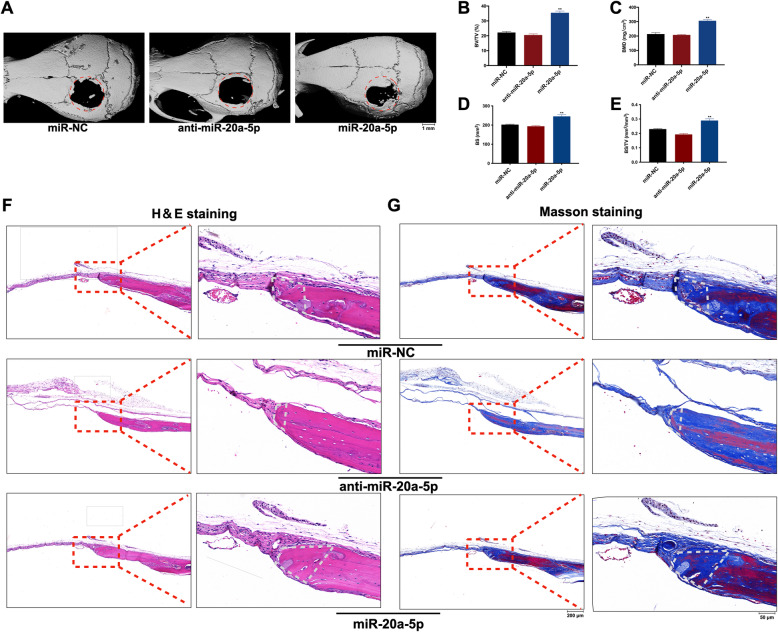
Fig. 3.
miR-20a-5p promoted the regeneration of calvarial defects. A Micro-CT images showed the regeneration of each calvarial defect in miR-20a-5p, anti-miR-20a-5p, and miR-NC groups. B BMD in miR-20a-5p, anti-miR-20a-5p, and miR-NC groups. C BV/TV in miR-20a-5p, anti-miR-20a-5p, and miR-NC groups. D BS in miR-20a-5p, anti-miR-20a-5p, and miR-NC groups. E BS/TV in miR-20a-5p, anti-miR-20a-5p, and miR-NC groups. F H&E staining illustrated new bone formation in miR-20a-5p, anti-miR-20a-5p, and miR-NC groups. White trapezoid represented new formed bone. G Masson’s trichrome staining showed new collagen fibers in miR-20a-5p, anti-miR-20a-5p, and miR-NC groups. White trapezoid represented new formed bone. *p <0.05 and **p <0.01 compared with the miR-NC group

