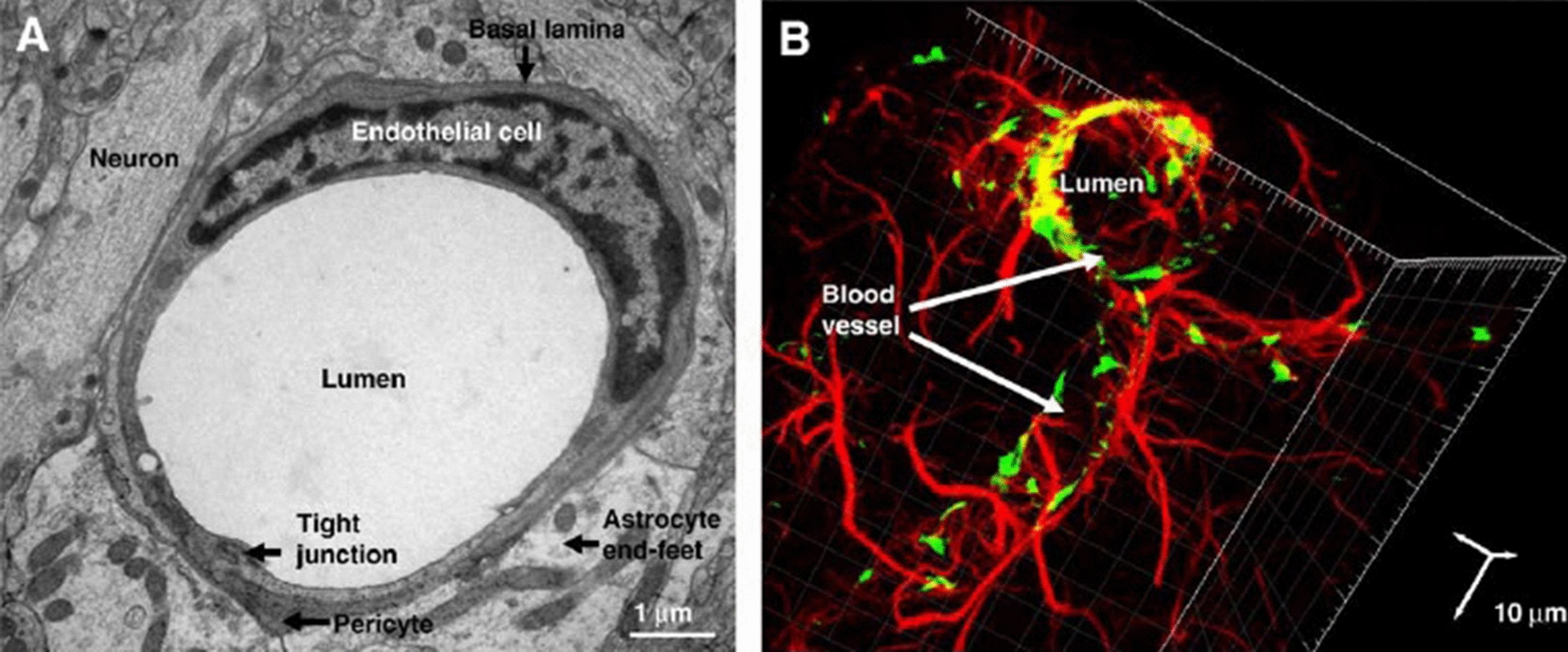
Fig. 1.

A Electron microscopy (TEM) of the neurovascular unit of rat brain section showing the vascular ECs, embedded pericytes in basal lamina and astrocytes, which are close to neurons. B Confocal microscopy imaging of cerebral vascular tree in rat brain section, Astrocytes (Red) surround endothelial cells (Green). Vascular ECs and astrocytes were stained for von- Willebrand factor (vWF) and glial fibrillary acidic protein (GFAP), respectively
(Adapted with permission [227]. Copyright 2008, Biochimica et Biophysica Acta (BBA)—Biomembranes)
