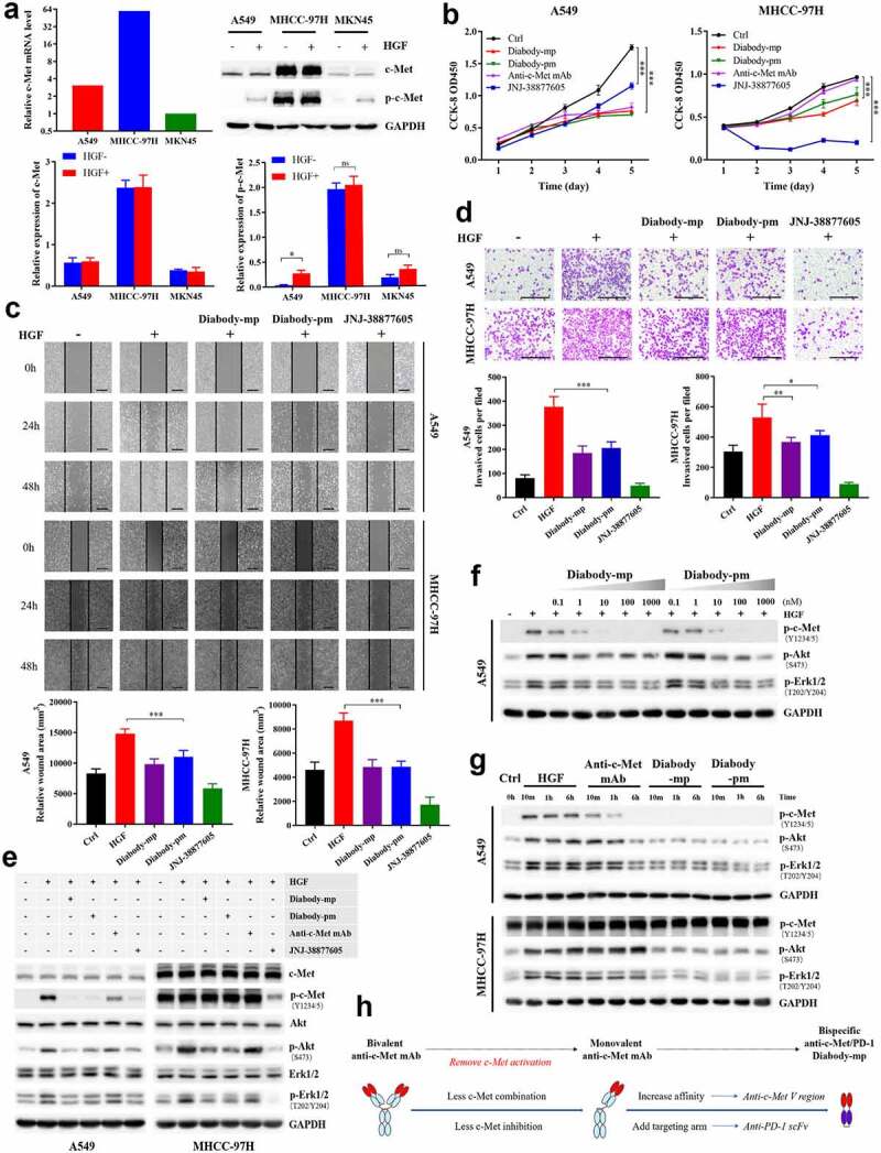
Figure 2.

Diabodies inhibit the proliferation and movement of tumor cells and the activation of c-Met signaling by antagonizing HGF binding to c-Met. a The mRNA levels of c-Met detected by quantitative reverse transcription polymerase chain reaction in A549, MHCC-97 H, and MKN45 cells. Expression level of p-c-Met (Y1234/5) and total c-Met protein levels in A549, MHCC-97 H, and MKN45 cells stimulated with or without HGF (1 nM). b A549 or MHCC-97 H cells treated with 100 nM of diabody-mp, diabody-pm, anti-c-Met mAb, or JNJ-38877805 and stimulated with 1 nM HGF were subjected to a Cell Counting Kit-8 assay, and viable cells were measured on days 1–5. c Representative images of A549 cells and MHCC-97 H cells incubated with 100 nM of diabody-mp, diabody-pm, and JNJ-38877605 (positive control) from HGF-induced wound healing assay; scale bar, 400 µm. d Representative images of A549 and MHCC-97 H cells incubated with 100 nM of diabody-mp, diabody-pm, and JNJ-38877605 (positive control) from HGF-induced transwell assay; scale bar, 400 µm. e Western blot analysis of the activation of c-Met downstream molecules in A549 or MHCC-97 H cell lysates. Cells were treated with 100 nM of diabody-mp, diabody-pm, anti-c-Met mAb, and JNJ-38877605 for 2 hr and analyzed by immunoblotting with the indicated antibodies. HGF (1 nM) was added to stimulate cells at 37 ℃ for 15 min before sample collection. f Diabody-mp or diabody-pm inhibits HGF-induced c-Met signaling activation in a dose-dependent manner. A549 cells were incubated with the indicated concentration of diabody-mp or diabody-pm at 37 ℃ for 2 hr. g Western blot analysis of c-Met and other phosphorylated-targets in A549 and MHCC-97 H cells lysates. The cells were treated with HGF, anti-c-Met mAb, diabody-mp, and diabody-pm at indicated time point. h Design ideas of diabody-mp
