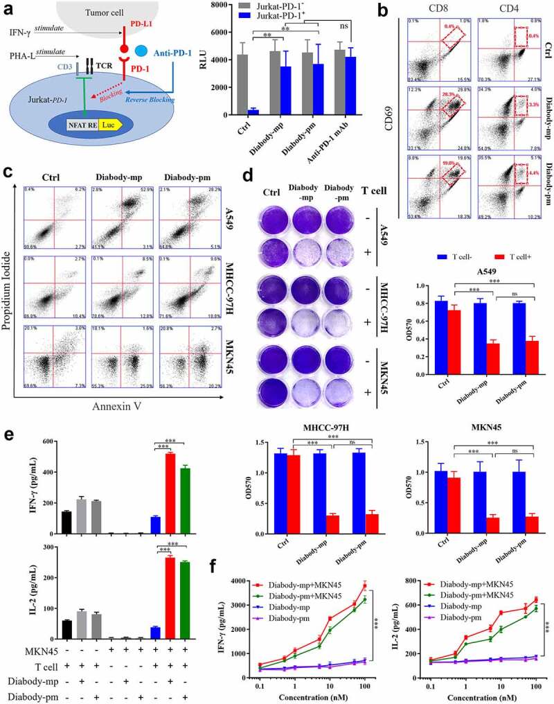
Figure 3.

Diabodies block the PD-1 pathway, mediating the cellular cytotoxicity of c-Met-positive tumor cells through T cell engagement. a Schematic diagram of a reporter gene assay to measure the anti-PD-1 activity of diabody-mp or diabody-pm (left). The levels of luciferase activity were determined (right). b FACS quantification of CD69+-activated CD8+ T cells and tumor-infiltrating CD4+ effector T cells obtained from a patient with HCCs and incubated with 100 nM of diabody-mp, diabody-pm, and anti-PD-1 mAb for 48 hr after digestion with enzymes. c Apoptosis detection in tumor cells co-cultured with activated human T cells and treated with 100 nM of diabody-mp or diabody-pm for 48 hr at an E:T ratio of 10:1. The percent of positive apoptotic cells is shown. d A549, MHCC-97 H, or MKN45 cells co-cultured with activated human T cells at an E:T ratio of 10:1 upon the addition of 100 nM diabody-mp or diabody-pm for 48 hr, next subjected to crystal violet staining. e IFN-γ and IL-2 released in the presence of MKN45 cells and T cells at an E:T ratio of 10:1 upon the addition of 100 nM diabody-mp or diabody-pm, as measured by enzyme-linked immunosorbent assay (ELISA) kit after 48 hr. f IFN-γ and IL-2 released in the presence or absence of MKN45 cells at an E:T ratio of 64:1 upon the addition of diabody-mp or diabody-pm, as measured by ELISA kit after 48 hr
