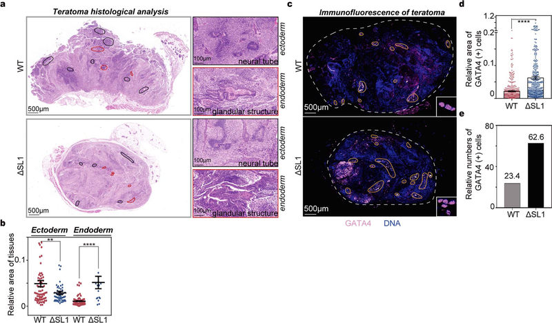
Extended Data Fig. 7 |. miRNA dosage control impacts germ layer specification during teratoma formation in mouse.
a, Histology of teratomas stained with hematoxylin and eosin (HE). Left: low power view of teratoma, differentiated areas are shown with different colors (ectoderm: marked with black; endoderm: marked with red). Right: representative images of ectoderm (neural tube) and endoderm (glandular structure) tissues. b, Immunofluorescence (IF) of GATA4 proteins (endoderm marker) in sections of teratomas derived from WT and ΔSL1 mESCs. Fluorescence signals of GATA4 are shown in pink, and DAPI stain is shown in blue. c, Comparison of the relative area of GATA4 (+) cells in teratomas derived from WT and ΔSL1 mESCs according to the immunofluorescence of 34 (WT:17, ΔSL1:17) sections of 18 (WT:9, ΔSL1:9) teratomas from 9 mice. d, The bar plot showing the GATA4 (+) cell numbers relative to the total area in teratomas derived from WT and ΔSL1 mESCs according to the staining in b.

