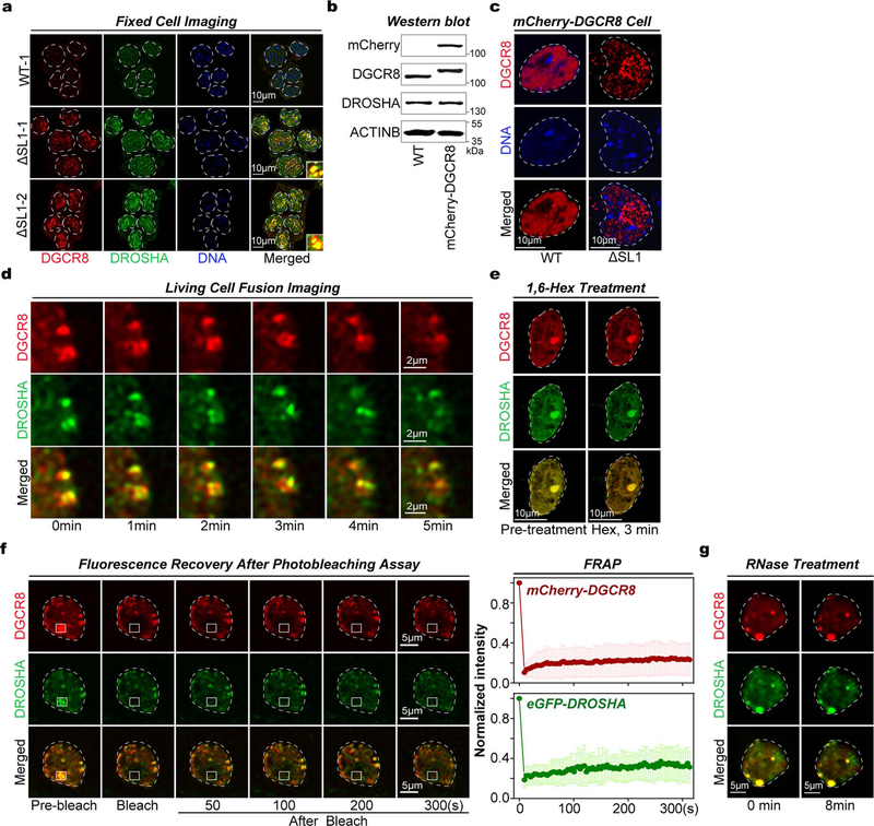
Extended Data Fig. 2 |. SL1 depletion drives irreversible Microprocessor aggregation in mouse ESCs.
a, Immunofluorescence (IF) followed by confocal imaging of DGCR8 and DROSHA proteins in WT and ΔSL1 mESCs. Fluorescence signals of DGCR8 and DROSHA are shown in red and green, respectively, and DAPI stain is shown in blue. The merged signals are also shown. b, Western blot of DGCR8 and DROSHA proteins in WT and reporter mESCs expressing endogenous mCherry-DGCR8 fusion protein. c, Representative time-lapse images of two proximate assemblies of Microprocessor in living ΔSL1 mESCs transfected with plasmids expressing tagged mCherry-DGCR8 and eGFP-DROHSA. d, Representative images of fluorescence recovery after photobleaching (FRAP) analysis of Microprocessor aggregates in living ΔSL1 mESCs transfected with plasmids expressing tagged mCherry-DGCR8 and eGFP-DROHSA. Targeted region is highlighted in a white box, and DGCR8 (red), DROSHA (green) and merged (yellow) signals are shown. See also Fig. 1g. e, Images of Microprocessor aggregates in ΔSL1 mESCs transfected with plasmids expressing tagged mCherry-DGCR8 and eGFP-DROSHA before and after microinjection with RNase.

