
Figure 1 .
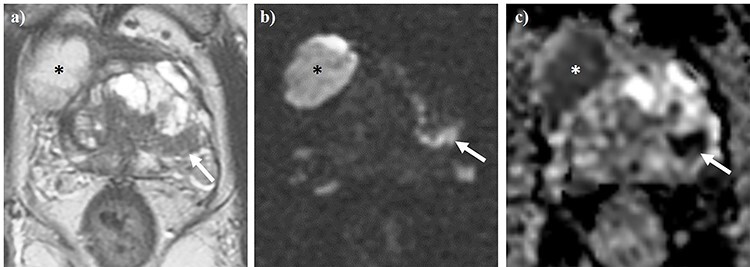
Axial T2WI (a) and diffusion-weighted imaging (b), alongside apparent diffusion coefficient (ADC) mapping (c) of the pelvis. The white arrow denotes a PI-RADS 5 lesion located in the left peripheral zone. Asterisks demonstrate one of the locules of a concurrent giant multilocular prostatic cystadenoma.