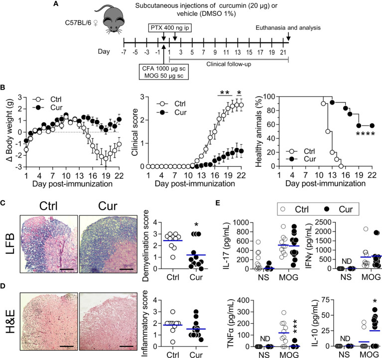
Figure 4.
Subcutaneous microdose curcumin is also neuroprotective. (A) WT C57BL/6 female mice were treated subcutaneously in the hind footpads every other day with curcumin (Cur, 10 µg each footpad, 20 µg per mouse, approximately 0.8 mg/kg) or vehicle (DMSO 1%; Ctrl), 7 days prior EAE induction (day 0) and until the end of the experiment, as shown. Clinical follow-up was performed daily, and after euthanasia (day 22), samples were collected to perform histopathological analysis and immunological tests. (B) Changes in body weight (left), clinical score (center) and percentage of healthy animals (right) were recorded daily after immunization (see Materials and Methods for details). Data is presented as the mean ± SEM (left and center). Percentage of healthy animals (right) was plotted as a Kaplan-Meier curve. (C, D) Representative images of the lumbar cord sections stained with (C) LFB and (D) H&E. Plotted data represents the (C) demyelination score and (D) inflammatory score obtained after image analysis from each animal in both treatment groups. Scale bar=25 µm. Each point represents individual animals and bars represent the mean of the group. (E) 2.0×10⁶/mL splenocytes were stimulated with MOG (20 µg/mL) or PBS (NS) for 72 h. The amounts of the indicated cytokine produced by cells in response to stimuli were quantified in supernatants by ELISA. ND, not detected. Results are plotted as individual animals and mean (n=10-12). *P < 0.05; **P < 0.01; ***P < 0.001; ****P < 0.0001. Statistical tests were: (B, left and center) Two-way ANOVA for repeated measures with Bonferroni’s post-test, (B, right) Mantel-Cox test and (C–E) Mann-Whitney U test.

