FIGURE 1.

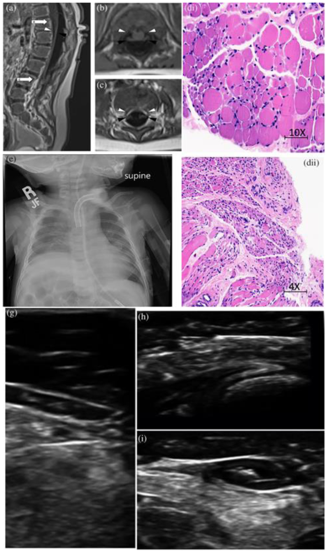
(a) Patient 1 MRI of the spine in sagittal view. Note marked volume loss of ventral cauda equina (white arrowhead) and intact dorsal cauda equina (black arrowhead). White arrows signify the spinal level of images (b) and (c) that show marked volume loss of ventral cauda equina (white arrowhead) and intact dorsal cauda equine (black arrowhead) in axial view. (d) Patient 1, hemotoxylin and eosin stain of quadriceps muscle with marked variation in fiber size with small group and fascicular atrophy typical of SMA1; (e) Patient 2, chest X-ray with elevation of Right hemi-diaaphragm; (f) Patient 2, hemotoxylin and eosin stain of muscle with variation in muscle size, rare myophagocytosis, regenerating fibers (g), (h), and (i) Patient 3, muscle ultrasound with severe atrophy and neurogenic changes of distal > proximal muscles
