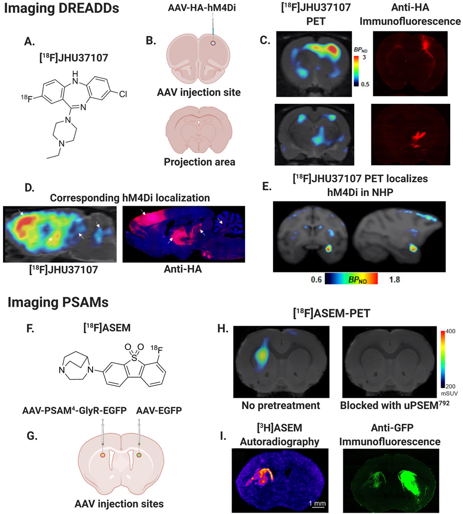
Fig. 3. PET-reporters for in vivo mapping of chemogenetic receptor expression.
A–E. Mapping DREADDs in rats and NHPs (modified from Bonaventura et al., 2019): A.) Chemical structure of DREADD radioligand [18F]-JHU37107. B.) AAV-HA-hM4Di injection site in rat right motor cortex (M1) and non-injected projection area. C.) Left: [18F]-JHU37107 PET reveals hM4Di expression at M1 injection site and putative projection sites, Right: Histological confirmation of hM4Di expression with hemagglutinin (HA) antibody immunofluorescence. D.) Anatomical localization of hM4Di from [18F]-JHU37107 PET in rats coincides with histological expression patterns of the HA-tagged DREADDs, arrows show corresponding anatomical sites of right M1 and motor cortical projection areas. E.) Localization of hM4Di in rhesus nonhuman primate (NHP) right amygdala with [18F]-JHU37107. F–I. Mapping PSAMs in mice (modified from Magnus et al., 2019): F.) Chemical structure of FDA approved radiotracer [18F]-ASEM capable of imaging PSAMs. G.) Schematic of AAV injection sites in mouse left dorsal striatum (AAV-PSAM4-GlyR-EGFP) and right dorsal striatum (AAV-EGFP control). H.) Left: [18F]-ASEM-PET showing left striatal PSAM4-GlyR expression, Right: [18F]-ASEM blocked following uPSEM792 pretreatment (1 mg/kg). I.) Left: Autoradiography of [3H]-ASEM binding in left dorsal striatum, Right: Immunofluorescence with anti-green fluorescent protein (anti- GFP) antibody showing PSAM4-GlyR-EGFP expression in left striatum and EGFP control in right striatum. Lack of PET and autoradiography binding in right striatum demonstrates specificity of ASEM for PSAM4-GlyR.

