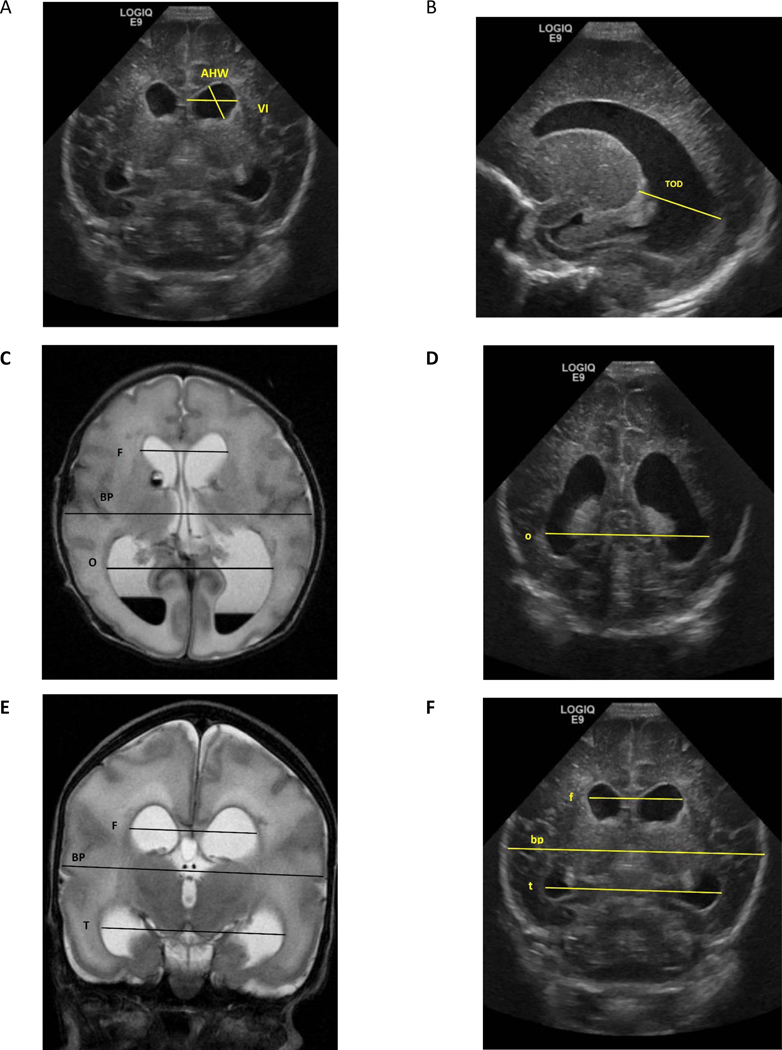
Figure 1: Demonstration of different ventricular measures and ratios.
Images from cUS and brain MRI of one-week old boy born at 30 weeks of gestation. A and F= Coronal cUS at level of Foramen of Monro, B= Parasagittal cUS, C= Axial View of T2 MRI. D= Coronal cUS at level of occipital horn E= Coronal View of T2 MRI, VI= Ventricular Index, AHW= Anterior Horn Width, TOD= Thalamo-Occipital Diameter MRI dimensions are in Capital letters= F= bifrontal horn, BP= biparietal, T= bitemporal horn, O =bioccipital horn
cUS dimensions are in small letters= f= bifrontal horn, bp= biparietal, t= bitemporal horn, o =bioccipital horn
Evans Ratio= F/BP by MRI or f/bp by US
Frontal and Temporal Horn Ratio= (F+T/2)/ BP by MRI or (f+t/2)/bp by cUS
Frontal and Occipital Horn Ratio (FOHR)= (F+O/2)/BP by MRI or (f+o/2)/bp by cUS

