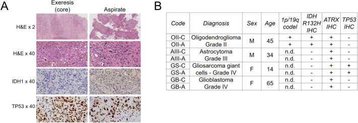
Fig 1. The diagnosis of glioma aspirates is highly similar to their corresponding core resections.
A, Representative immunohistochemistry images of the gliosarcoma of giant cells used in this study. H&E, haemotoxylin and eosin. B, Diagnosis of the samples used in the present study. C = tumour core resection. A = cavitational ultrasonic surgical aspirate. Age = Age at diagnosis.

