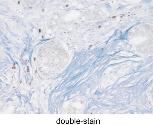
Fig 8. Tumor-infiltrating lymphocytes and collagen fibers using double-staining.

Detection of tumor-infiltrating lymphocytes (brown) and collagen fibers (blue) double-stained using immunohistochemical staining and Masson’s trichrome staining (x200 HPF).
