Fig. 1.
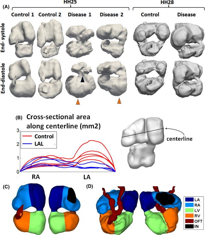
a Samples of reconstructions of the chick embryonic hearts blood space (other reconstructions are shown in supplementary Figs. 1 and 2). At HH25, there was only one atrio-ventricular junction, but by HH28, the atrio-ventricular junction divided into two. Left atrial ligation severely reduced left atrial volume, caused the ventricle to adopt a more triangular shape with a sharper apex (black arrow), and medially shifted the atrioventricular junction in some hearts (orange arrow, LAL1). b Cross-sectional area of the atrium along medial–lateral direction, during atrial end-diastole, demonstrating narrowing in the left atrium but not in the right atrium. c-d Delineation of the embryonic heart into the four cardiac structures for the c HH25 and d HH28 hearts. RA-right atrium, LA-left atrium, RV-right ventricle, LV-left ventricle, OFT-outflow tract, IN-sinus venosus inlet

