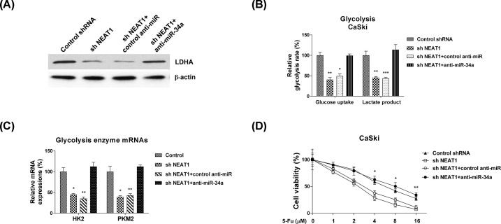
Figure 8. The NEAT1-promoted 5-Fu resistance is through miR-34a/LDHA axis.
(A) CaSki cells were transfected with control shRNA, NEAT1 shRNA alone, NEAT1 shRNA plus control antisense, or NEAT1 shRNA plus miR-34a antisense for 48 h, the LDHA protein expressions were detected by Western blot. β-actin was a loading control. (B) The above cells were subjected to glucose uptake and lactate product measurements. (C) The mRNA levels of glycolysis enzymes were detected by qRT-PCR from the above transfected cells. GAPDH was used as an internal control. (D) The above transfected CaSki cells were treated by 5-Fu at 0, 1, 2, 4, 8 or 16 uM for 48 h. Cell viabilities were examined by MTT assay. Experiments were performed in triplicate. All data were shown as mean ± S.D. *, P<0.05; **, P<0.01; ***, P<0.001.

