
Figure 4.
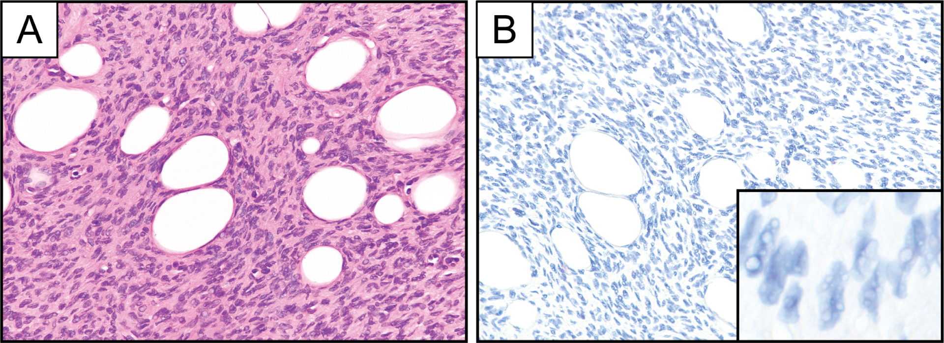
Representative photomicrographs of hematoxylin and eosin stain (A) and PDGFB RNA chromogenic in situ hybridization (B; PDGFB-1 probe set) in a case of PDGFD-rearranged DFSP. Inset represents PDGFB RNA chromogenic in situ hybridization at high magnification.