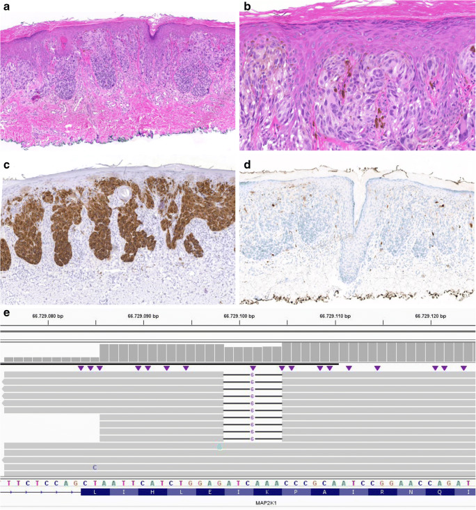
Fig. 1.
Microscopical and molecular findings case 2. a H&E, × 5: relatively symmetrical, compound melanocytic lesion extending into the superficial dermis with large nests at the junction. b H&E, × 40: epithelioid to spindle-shaped melanocytes with pale, eosinophilic cytoplasm. Variation in size and shape of the cells was encountered with large nuclei with prominent nucleoli and nuclear vacuoles. A few multinucleated cells were seen, c Melan A stain, × 5: diffusely positive. d p16 stain, × 5: negative in the majority of the lesion. e Integrative Genomics Viewer (IGV) visualization of the sequence data containing MAP2K1 in-frame deletion c.307_312del (p.Ile103_Lys104del) in 24% of the reads (RefSeq NM_002755.3). The black lines indicate the location of the deletion

