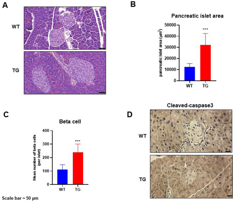
Figure 5.
Pancreatic islet architecture. The pancreatic sections were prepared and stained with hematoxylin and eosin (H&E) or anti-cleaved caspase-3 antibody. (A) Representative images of H&E-stained pancreatic sections from different groups. Scale bar = 50 µm. (B,C) Pancreatic islet area and beta cell number were measured as described in the methods section. Data are shown as mean ± standard error of mean. ***p < 0.01. (D) Immunohistochemical analysis of cleaved caspase-3 in the pancreas islets. Scale bar = 50 µm.

