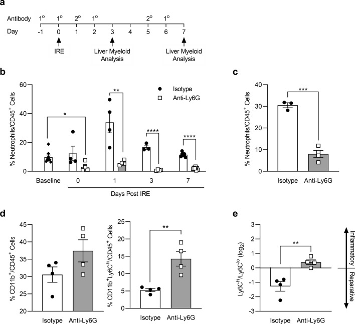
Figure 5.
Neutrophil depletion increases the proportion of CD11b+Ly6Chi cells in the liver after IRE and shifts the macrophage population toward a more pro-inflammatory state. (a) Neutrophil depletion protocol and timeline in which rat IgG2a (isotype control) or anti-Ly6G were given as 1° antibodies and anti-rat IgG given as the cross-linking 2° antibody. The timing of IRE treatment and analysis of liver myeloid cells are indicated by arrows. (b) Percentage of neutrophils in the blood at baseline (prior to any IRE or antibody treatments), and on days 0, 1, 3, and 7 after IRE treatment with the neutrophil depletion regimen shown in (a). (c) Percentage of neutrophils in IRE-treated liver parenchyma 3 days after treatment in mice given isotype control or anti-Ly6G. (d) Total CD11b+ cells or CD11b+Ly6Chi cells as a percentage of CD45+ leukocytes in IRE-treated liver parenchyma 7 days after treatment in mice with or without neutrophil depletion. (e) Ly6Chi to Ly6Clo ratio in IRE-treated liver 7 days after treatment in mice given isotype control versus anti-Ly6G. For all graphs, unpaired two-tailed t tests show *p < 0.05, **p < 0.01, ***p < 0.001, and ****p < 0.0001. Data points represent individual animals in each condition and/or timepoint (n = 3–8). Graph bars and error bars show mean ± SEM.

