Figure 6.
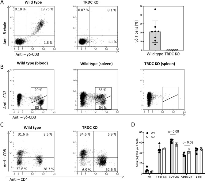
No γδ T cells are present in blood and spleen of TRDC-knockout pigs. (A) PBMC of TRDC-knockout and wild type pigs were stained with anti-CD3, anti-δ chain and anti-γδ T cell-specific CD3 mABs. CD3 positive cells were gated and δ chain vs. γδ T cell-specific CD3 dotblots are displayed. About 20% of peripheral blood T cells were γδ T cells in wild type pigs while no γδ T cells were detected in TRDC-knockout pigs. (B) PBMC and spleen cells were isolated and staind for CD2 and γδ T cell-specific CD3. Typically in wild type pigs the majority (80%) of γδ T cells in the blood belong to the CD2 negative phenotype (left dot blot), while in the spleen the majority (66%) of γδ T cells are CD2 positive (middle dot blot). No γδ T cells of either type were detected in the spleen of TRDC-knockout pigs. (C) Most γδ T cells are CD4 and CD8 negative. Accordingly TRDC-knockout pigs have minimal numbers of CD4/CD8 double negative T cells. (D) The percentage of lymphocyte subpopulations of all lymphocytes without γδ T cells were compared of wild type and TRDC-knockout pigs. No significant difference of any lymphocyte subpopulation was detected.

