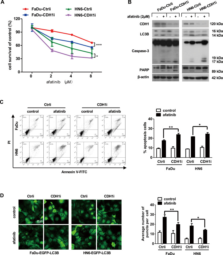
Fig. 6. Afatinib induces more apoptosis and less autophagy in HNSCC cells with CDH1 depletion.
A FaDu and HN6 cells with CDH1 depletion as well as the corresponding control cells were treated with the indicated concentration of afatinib for 24 h. Cell viability was examined by SRB assay. Statistical differences between the two groups were evaluated by two-way ANOVA. *p < 0.05, ***p < 0.001. B, C FaDu and HN6 cells with CDH1 depletion as well as control cells were treated with 2 μM afatinib for 24 h. The levels of CDH1, LC3B, and apoptosis-associated proteins caspase-3 and PARP were measured by western blot analysis (B), while flow cytometry analysis was carried out to evaluate apoptosis (C). D FaDu and HN6 cells stably expressing EGFP-LC3B were treated with 2 μM afatinib for 24 h. Cells were then fixed, imaged, and EGFP-LC3B fluorescent spots were quantified. Scale bar, 25 µm. The data are presented as mean ± SD from three independent experiments. *p < 0.05, **p < 0.01, ***p < 0.001.

