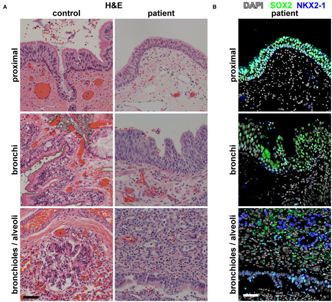
Figure 2.
Histological and molecular analysis of the identity of the broncho-esophageal fistula and more distal airway. (A) H&E staining of the patient vs. control tissues from the “proximal” epithelium (broncho-esophageal fistula vs. trachea, respectively), bronchi, and distal bronchioles and alveoli. (B) Immunofluorescence staining of patient tissue for transcription factors SOX2 and NKX2-1 in sections from the proximal epithelium (broncho-esophageal fistula vs. trachea, respectively), bronchi, and distal bronchioles and alveoli. Scale bars represent 50 μm.

