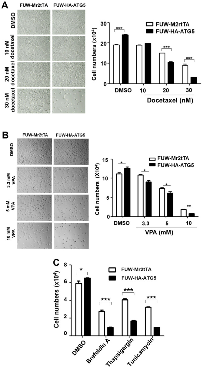
Figure 4.
Restoration of ATG5-dependent autophagy in DU145 cells sensitizes them to the cytotoxicity of chemotherapeutic drugs and to ER stressors. (A) Light phase microscopic images of cells treated with the indicated concentrations of docetaxel for 72 h (left panel; magnification, ×200) and semi-quantification of the inhibition of proliferation by docetaxel treatment in ATG5-expressing or vector control cells (right panel). (B) Light phase microscopic images of the cells treated with the indicated concentrations of VPA for 48 h (left panel; magnification, ×200) and semi-quantification of the inhibition of proliferation by docetaxel treatment in ATG5-expressing or vector control cells (right panel). (C) Semi-quantification of the inhibition of proliferation by the ER stressors brefeldin A (500 nM), tunicamycin (2.0 µg/ml) and thapsigargin (250 nM) for 24 h in the ATG5-expressing or the vector control cells. In all the quantification figures, the data were from three independent experiments. *P<0.05; **P<0.01; ***P<0.001. ATG5, autophagy related 5; VPA, valproic acid.

