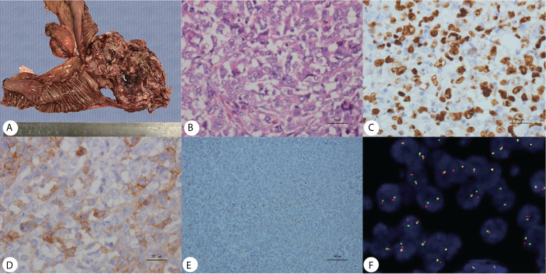
Figure 3.
(A) Gross pathology revealed an ileum tumor specimen with brittle texture and multiple ulcerations on the surface; (B) H&E staining revealed small round blue cells; ×40. (C–E) Immunohistochemistry showed positive Ki67, CD99 and Fli-1 staining; ×40. (F) Molecular analysis revealed positive EWSR1 fusion genes.

