
FIGURE 1.
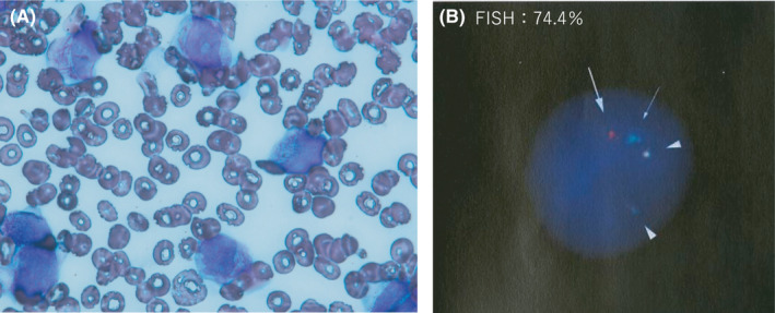
Acute promyelocytic leukemia at diagnosis. A, Bone marrow smear, (B) FISH (Fluorescence in situ hybridization) analysis of metaphase spreads and interphase nuclei of bone marrow cells, a signal of PML (15q22) probe (arrow), a signal of RARA (17q21) probe (thin arrow), two signals of PML/RARA probe (arrowhead) G‐banded karyotype of bone marrow cells: 46, XY, t(15;17)(q22;q21)[8]/46,XY[12]