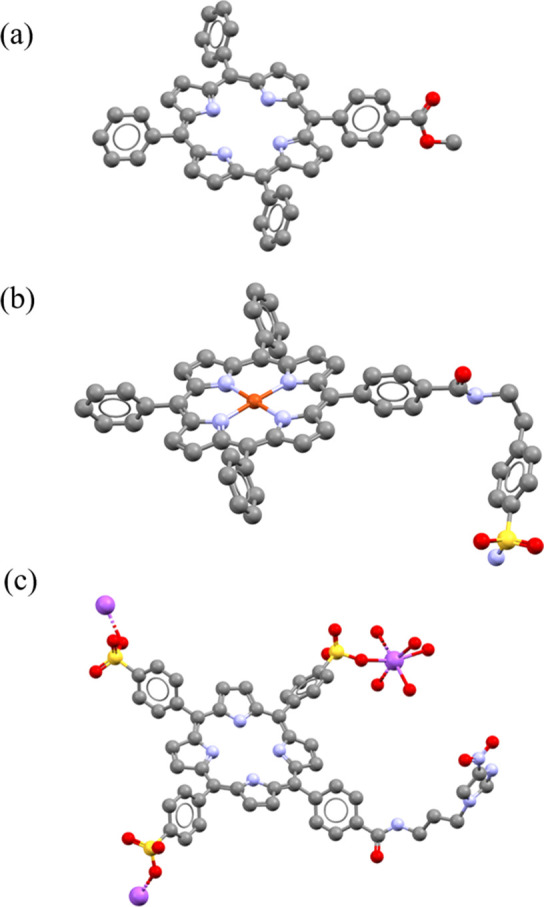
Figure 4.

Molecular structures determined from single crystal X-ray diffraction for compounds 2 (a), 3.17 (b), and 3.22 (c). Color code: magenta = Na, blue = N, yellow = S, red = O, gray = C. H atoms and disordered units have been removed. Crystallography details for all structures are given in the SI.
