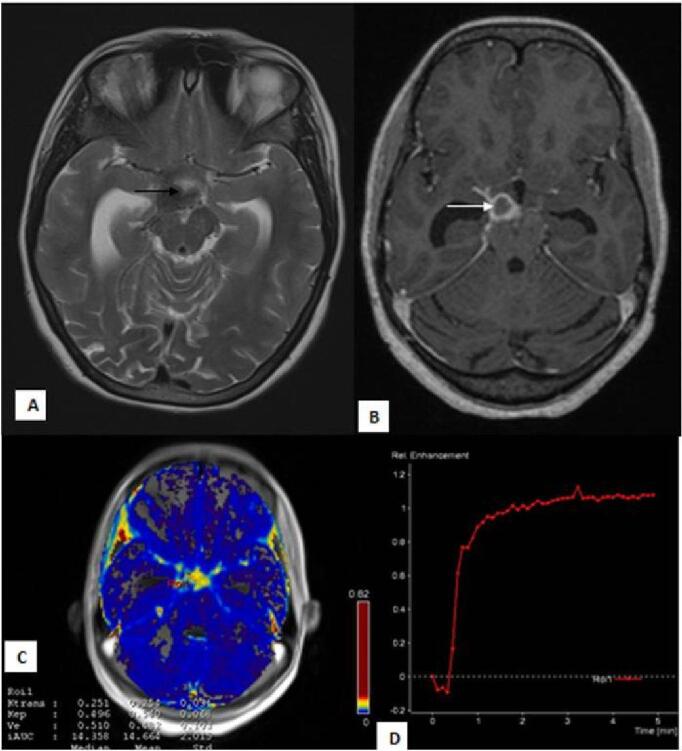
Fig. 2.

Perfusion changes in patient with “enhancing” basal exudates: 21-year female presented with complaints of headache and signs of meningeal irritation. T2W ( A ) image shows dilated temporal horns of bilateral lateral ventricles with hypointense exudates in the suprasellar cistern ( black arrow ). T1 post contrast ( B ) image shows thick basal exudates involving the suprasellar cistern ( white arrow ). Physiological map ( C ) shows (mean K trans 0.254 minute –1 and mean Ve 0.482) high permeability of contrast in the cistern. Signal intensity-time curve ( D ) shows increased intensity above the baseline.
