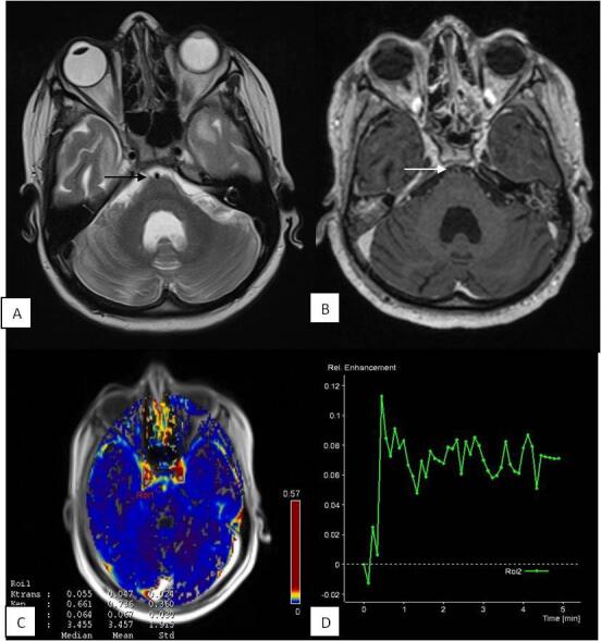
Fig. 3.

Perfusion changes in patient with “non-enhancing” basal cisterns: 35-year male presented with headache, fever, and altered sensorium. CSF PCR was positive for Mycobacterium tuberculosis . T2W ( A ) image shows normal appearing prepontine cistern ( black arrow ). T1 post contrast ( B ) image shows no enhancement in the prepontine cistern ( white arrow ). Physiological map ( C ) shows (mean K trans 0.047 minute –1 and mean Ve 0.067) low permeability of contrast in the cistern. Signal intensity–time curve ( D ) of the cistern shows reduced intensity with flattened baseline. CSF, cerebrospinal fluid; PCR, polymerase chain reaction.
