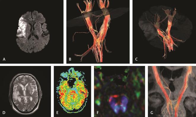
Fig. 3.
( A–G ) Case 3: A 64-year-old male with left-sided hemiplegia (NIHSS19). Imaging at 10 hours of stroke: (A) diffusion-weighted image reveals right temporal acute infarction. (B and C) 3D reconstruction of corticospinal tract reveals displacement, mild attenuation, interruption at the motor cortex and corona radiata, decreased fiber number of ipsilateral CST. Imaging at 30 days of stroke: (D) axial T2 WI reveals a decrease in the size and intensity of infarction. (E) Axial FA map at the pons reveals a marked decrease in FA on the right side (rFA: 0.83) denoting Wallerian degeneration. (F) Color-coded map at the pons reveals distortion of right CST. (G) 3D reconstruction of both CSTs reveals attenuation and a decreased number of fiber of CST at the pons denoting Wallerian degeneration. CST, corticospinal tract; FA, fractional anisotropy; NIHSS, National Institute of Health Stroke Scale; rFA, fractional anisotropy ratio; WI, weighted image.

