
Fig. 1.
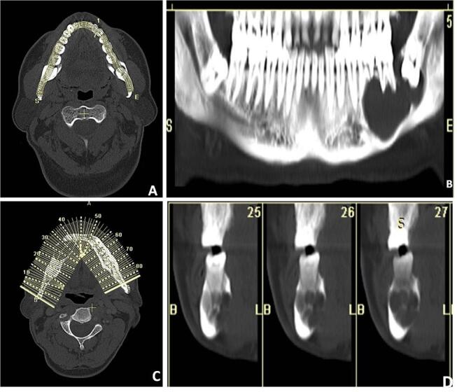
( A–D ) Panoramic reconstruction: the images are reconstructed using a dental computed tomography (CT) application where multiple dots are deposited along the curve of the jaw starting from one end of the jaw to the other end ( A ). Then the program connects these dots to form a smooth curved line that is superimposed to form a panoramic image ( B ). Orthoradial reconstruction. The same dental CT application can also be used to create para-axial views where the application creates multiple perpendicular lines along the smooth curved line ( C ) to give a series of paraxial views along each corresponding tooth ( D ).